
CARDIAC PHASED ARRAY PROBE IMAGE DEGRADATION

- Crystals are grouped closely together.
- Sound waves originates from a single point and fan outward,creating a sector-type Image.
- Smaller and flatter footprint them the curvilinear probe.
- Probe has fequency between 2-8MHz.
- Currently used for cardiac Imaging,Imaging between ribs and small spaces.
The issue is almost certainly caused by a significant knock to the head of the probe as can be seen in the image below.

Now if we compare a good ultrasound image from the same phantom on the same ultrasound system. Firstly the good image, you can see how clear the phantom dots throughout the image are:

Now comparing with an image taken from the faulty probe:
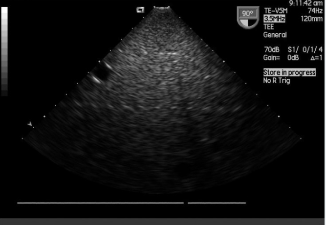
With such gradual deterioration, it is sometimes difficult to recognize the problem as it just gets more and more difficult to scan patients.


No comments:
Post a Comment